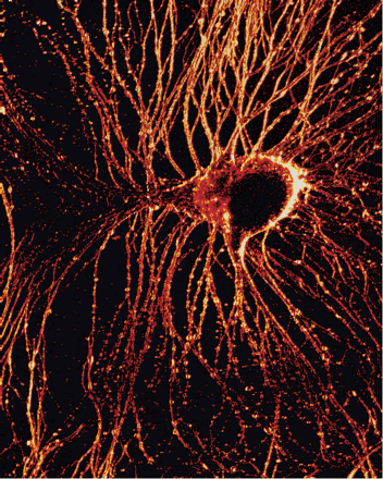
Second-harmonic generation microscopy image of a primary cultured Aplysia neuron stained with the membrane dye DHPESBP. The signal is modulated by membrane potential and was found to be capable of recording action potentials with 0.6 μm and 0.833 msec spatiotemporal resolution. The high-resolution and deep tissue imaging capability of this nonlinear microscopy technique should prove valuable to future electrophysiology studies.
Technique
Second harmonic generation microscopy
Citation
Optical Recording of Action Potentials with Second-Harmonic Generation Microscopy, Daniel A. Dombeck et al., The Journal of Neuroscience, January 28, 2004, 24(4):999–1003; doi:10.1523/JNEUROSCI.4840-03.2004
