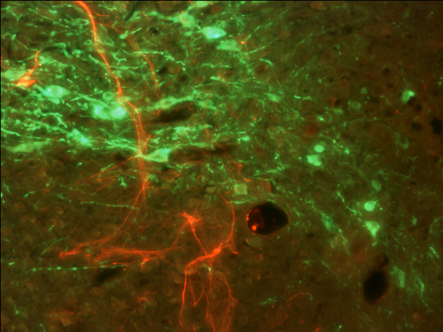
[Permanent link] Immunofluorescent colocalization of Oxytocin and Tyrosine Hydroxylase in the Parventricular Nucleus of a steroid treated hamster
Zoom: 1× 2× Fit monitor
Remove cookie
To enable HTML5 features, use a modern, JavaScript-enabled browser such as Google Chrome or Mozilla Firefox!
Labeling of the millions of axons crossing the internal capsule of the mouse embryo. Red: neurofilament 165kD staining. Green: EGFP from Ngn2 EGFP-KI knockin mice provided by Dr Francois Guillemot. Horizontal sections.
Melloni Agression Lab, Department of Psychology, Northeastern University, Boston USA
